

0 Views
Department of Entomology, ANGRAU-S.V. Agricultural College, Tirupati-517 502.
A promising alternative for insect pest control in agriculture is the use of entomopathogenic fungi. The objectives of this work were to isolate and identify strains of entomopathogenic fungi from brown plant hopper, Nilaparvatha lugens in rice ecosystems of Nellore and West Godavari districts of Andhra Pradesh. Two native strains of entomopathogenic fungi were isolated from the infected BPH cadavers. According to their macroscopic and microscopic traits, one isolate was identified as Beauveria bassiana species (coded as Bb-NLR) and another isolate was identified as Metarhizium anisopliae species (coded as Ma-NLR). Bb-NLR was isolated from a BPH cadaver collected from the Agricultural Research Station (ARS), Nellore, while Ma-NLR was isolated from a BPH cadaver collected from Chinnacherukuru village in T.P. Gudur mandal of Nellore district. No entomopathogenic fungi were recovered from soil samples despite multiple isolations. These native strains offer potential for development of biocontrol agents targeting major rice insect pests.
KEYWORDS: Entomopathogenic fungi, brown planthopper, nilaparvathalugens, morphological identification.
Rice (Oryzasativa L.) is a vital food crop globally, serving as the staple diet for over half of the world’s population. It plays a crucial role in ensuring food security and provides a major source of livelihood and employment, particularly in developing nations such as India. Approximately 90 per cent of global rice cultivation and consumption occurs in Asia. However, sustaining productivity amidst increasing pest pressure remains a major challenge for farmers and agricultural scientists. Despite technological advancements, rice production continues to face significant challenges from both abiotic and biotic stresses. Insect pests remain a major biotic constraint, causing substantial yield losses and threatening crop sustainability. Among the insect pests, the brown planthopper (Nilaparvatalugens, Hemiptera: Delphacidae) has economic importance in rice ecosystems across Asia. The brown planthopper is a monophagous herbivore is recognized as one of the most destructive insect pests of rice in Asia, capable of causing 20–80 per cent yield losses through both direct and indirect damage (Balachiranjeevi et al., 2019). N. lugens causes damage by feeding on phloem sap and transmitting viral pathogens, including rice grassy stunt virus (RGSV) and rice ragged stunt virus (RRSV), resulting in significant physiological stress and hopperburn symptoms (Helina et al., 2019). However, indiscriminate chemical use for
the control of N. lugens has led to adverse consequences such as pest resurgence, resistance development, non-target effects and environmental contamination.These concerns have prompted the adoption of Integrated Pest Management (IPM), with an emphasis on ecologically sustainable approaches. Among these, biological control utilizing microbial agents has gained attention as an environmentally benign and effective alternative. Entomopathogenic fungi (EPF) are promising candidates for biological control due to their ability to infect and kill a wide range of insect pests. More than 700 fungal species, mainly from Deuteromycetes and Entomophthorales, are known to be entomopathogenic (Wang et al., 2020). Rice ecosystems, characterized by high humidity and a favorable microclimate, support the natural occurrence and activity of EPF. Species such as Beauveriabassiana and Metarhiziumanisopliae have demonstrated high virulence against key rice pests (Malekan et al., 2015). The successful application of EPF in field conditions often depends on the selection of locally adapted and highly virulent strains. Indigenous isolates tend to perform better under native environmental conditions (Jackson et al., 2000). In this context, the present study aims to isolate and identify indigenous entomopathogenic fungi associated with brown plant hopper in the rice ecosystem.
The study was conducted during the rabi season of 2024 – 2025 at the Agricultural Research Station, Nellore (14.4303ºN,79.9987ºE), with the objective of Isolation, identification of indigenous entomopathogenic fungi (EPF) associated with brown plant hopper in the rice. A systematic roving survey was carried out in rice fields of Nellore and West Godavari districts, Andhra Pradesh during October 2024 – February 2025. In total, 18 villages were selected across six mandals (three mandals per district, three villages per mandal). Naturally infected insect cadavers showing external mycosis symptoms were collected using sterile fine brushes into sterile glass vials by labelling them. Rhizospheric soil samples (top 5–10 cm) were also collected near cadaver sites using a sterile spatula, placed in sterile polyethylene bags and stored at room temperature (25 ± 2°C) until processing.
media and incubated for 7–14 days under sterile conditions to allow colony development. To obtain pure cultures, hyphal tips were subcultured repeatedly onto fresh agar media. The purified fungal isolates were maintained on agar slants and preserved in glycerol at –19°C for long-term storage and future experimentation.
Ten soil samples were collected from surveyed fields and 1 gram from each sample was used for serial dilution. The soil was initially suspended in 10 ml of sterile distilled water (autoclaved at 121°C, 15 psi for 15 minutes) to prepare a 10⁻¹ dilution, followed by successive 1 ml transfers into 9 ml sterile water to achieve dilutions up to 10⁻¹¹. From the 10⁻¹¹ dilution, 1 ml was aseptically transferred into sterile Petri plates inside a Laminar Air Flow chamber. Approximately 10–20 ml of molten and cooled Sabouraud’s Maltose Agar with Yeast Extract was
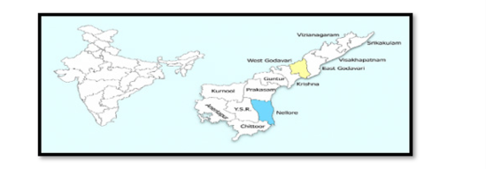
Fig. 1. Survey map showing collection of insect cadavers of BPH and soils from Nellore & West godavari districts of Andhrapradesh, India
The insect cadavers collected during the field survey were initially surface-sterilized by immersing them in 1 per cent sodium hypochlorite solution for 1 minute. This was followed by three successive rinses in sterile distilled water to remove any residual disinfectant. Following sterilization, the cadavers were transferred aseptically onto sterile Petri plates lined with moistened filter paper to maintain humidity. The plates were incubated at 25 ± 1°C for 3–5 days to facilitate the outgrowth of fungal mycelia from the cadaver surface.
Emerging fungal growth was carefully transferred to Sabouraud Dextrose Agar with Yeast extract (SDAY)
poured into the plates and gently swirled for even mixing before allowing it to solidify. The inoculated plates were incubated at 25°C ± 2°C for a duration of 10 days to facilitate fungal growth and isolation.
Fungal morphology was examined based on colony texture, mycelial structure,conidiophores, phialides, and conidia. Fungal mounts were prepared using lactophenol cotton blue stain on sterile slides and observed under a Phase Contrast Microscope (OLYMPUS BX43) at 40X &100X magnification. Identification was based on standard taxonomic keys and morphological traits.
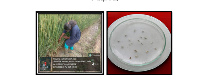
Fig. 2. Collection of insect cadavers of BPH from rice field in Nellore and west godavari districts of Andhra Pradesh
Survey efforts were focused in three mandals per district;Nellore Rural, Kodavaluru, and T.P. Gudur in Nellore district; and Penumantra, Achanta, and Palakollu in West Godavari district—covering different rice varieties and crop stages to enhance the possibility of capturing natural epizootics of entomopathogenic fungi in the field. In Nellore district brown planthopper cadavers were collected across all locations, mainly from rice varieties BPT 5204, KNM 1638, and MTU 1010 during tillering to grain filling stages. In West Godavari district, brown planthopper cadavers were predominantly found in MTU 7029 fields at the flowering stage. During the course of the survey, a total of 10 BPH cadavers suspected to be infected with entomopathogenic fungi were collected. The cadavers were collected based on visible signs of fungal infection under field conditions and brought to the laboratory for fungal isolation. Additionally, ten rhizospheric soil samples were also collected from the surveyed sites. However, none of the soil samples yielded entomopathogenic fungi despite repeated isolations using standard culturing techniques.
From the BPH cadavers collected from rice ecosystems, two fungal isolates showing typical characteristics of entomopathogenic fungi were isolated. Of these fungi, one isolate corresponded to Beauveria bassiana (coded as Bb-NLR) and one isolate corresponded to Metarhizium anisopliae (coded as Ma-NLR). Bb-NLR was isolated from a BPH cadaver collected from the Agricultural Research Station (ARS), Nellore, while Ma-NLR was identified from a BPH cadaver collected from Chinnacherukuru village in T.P. Gudur mandal of Nellore district. Remaining eight isolates were identified as plant pathogens morphologically under microscope.
The morphological identification (employing macroscopic and microscopic information) allowed the identification of the strains to the genera and species levels. In the present work, the fungal isolations of B. bassiana and M. anisopliae presented similar shapes, sizes and dispositions of conidia with the descriptions made by Samson et al. (1988), Humber (2005, 2012) and Rehneret al. (2005). All the fungal strains obtained corresponded to microorganisms of the Phylum Ascomycota, Sordariomycetes class, and the families Cordycipitaceae for Beauveria, and Clavicipitaceae in the case of Metarhizium (Humber, 2005, 2012).
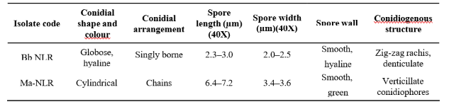
The isolate Bb-NLR exhibited white, powdery colonies with cottony to velvety aerial mycelium and a uniformly circular growth pattern. After 10–12 days of incubation at 25 ± 1°C, colonies attained a diameter of approximately 6.5 cm. Microscopic examination showed globose, hyaline conidia borne singly on a denticulated, zig-zag rachis, a characteristic feature of Beauveria bassiana. The conidia were smooth-walled, hyaline and measured 2.3–3.0 µm in length and 2.0–2.5 µm in width. Kirubhadharsini et al. (2017) also recorded the macroscopic characters of Beauveria isolates, which exhibited a white colony colour, powdery to cottony texture and a round shape and microscopic examination of Beauveria isolates revealed globose to sub-globose conidia with hyaline hyphae.
Table 1. Survey Locations and details of Entomopathogenic Fungi-Infected BPH Cadavers and soil collected from Rice Ecosystems of Nellore and West Godavari Districts (rabi 2024–2025)
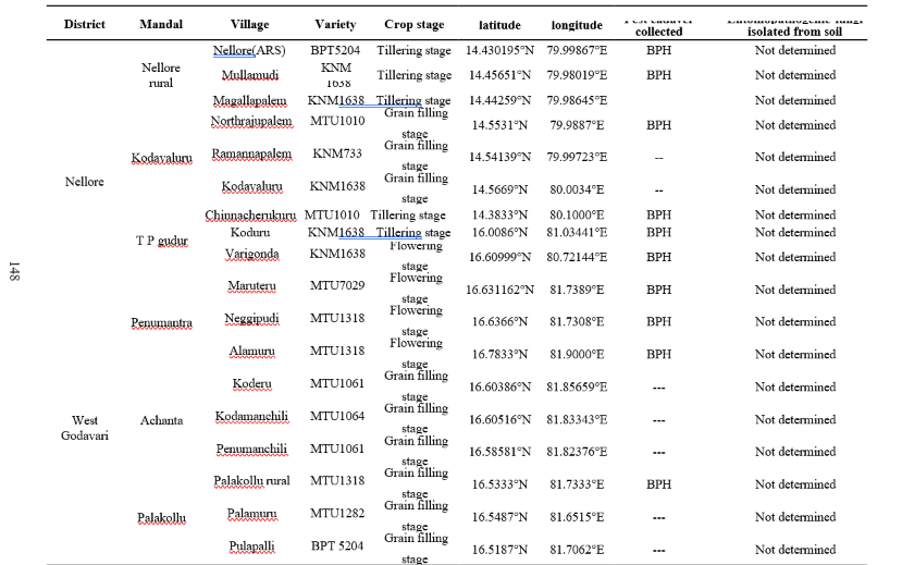
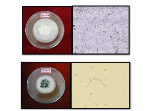
Fig. 3. Macroscopic and microscopic identification of fungal isolates (a) Morphology of Beauveria bassiana colony and (b) B. bassiana conidia (Bb-NLR) at 40X (c) Morphology of Metarhizium anisopliae colony (d) M. anisoplia econidia (Ma-NLR) at 40X
The Ma-NLR isolate displayed colonies with light to olive-green pigmentation, compact and granular in surface texture, and reached a diameter of approximately 5.8 cm after 10–12 days of incubation. Microscopic observation revealed cylindrical conidia arranged in chains on verticillate conidiophores. The conidia were smooth-walled, green and measured 6.4–7.2 µm in length and 3.4–3.6 µm in width. These morphological characteristics are consistent with previous reports for Metarhizium anisopliae, including the observations made by Elham et al. (2018), who reported similar traits on Potato Dextrose Agar. Bai et al. (2015) observed that colony borders of all the isolates of M. anisopliae were regular. During sporulation, the colour of colonies became yellow green or olivaceous green to dark green. The average spore width was 2.10- 4.10 μm, while the average spore length was 3.20- 7.69 μm. The spores were categorized as oval, round, or elongated based on their length and width.
The present work presents information of indigenous entomopathogenic fungi associated with brown plant hopper, N. lugens in rice ecosystem of Nellore district, Andhra Pradesh. Two native strains of entomopathogenic fungi were isolated from the infected BPH cadavers. According to their macroscopic and microscopic traits, one isolate corresponded to B. bassiana species and one isolate corresponded to M. anisopliae species. No entomopathogenic fungi were recovered from soil samples despite multiple isolations. The native isolates have potential for use in eco-friendly rice pest management strategies.
Table 2. Macroscopic characteristics of isolated entomopathogenic fungal isolates
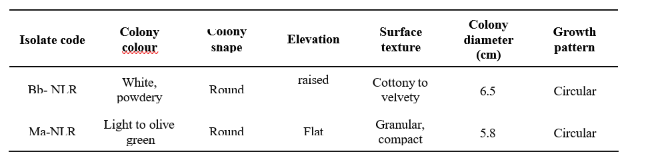
Table 3. Microscopic characteristics of isolated entomopathogenic fungal isolates
Bai,N.S.,Sasidharan,T.O.,Remadevi,O.K.,Dharmarajan, P., Pandian, S.K and Balaji, K. 2015. Morphology and RAPD analysis of certain potentially entomopathogenic isolates of MetarhiziumanisopliaeMetsch. (Deuteromycotina:Hypocreales). Journal of Microbiology and Biotechnology Research. 5 (1): 34- 40.
Balachiranjeevi, C.H., Prahalada, G.D., Mahender, A, Jamaloddin, M, Marfori- Nazarea, C.M., Vinarao, R, Sushanto, U, Baehaki, S.E., Li ZK and Ali, J. 2019. Identification of a novel locus, BPH38(t), conferring resistance to brown planthopper (Nilaparvata lugens Stal.) using early backcross population in rice (Oryza sativa L.). Euphytica .215(185):1–14.
Elham, M.S.H., Pong, K.K., Lee, G.E.L and Azmi, W.A. 2018. Occurrence of Entomopathogenic Fungus, Metarhizium anisopliae isolated from Island, BRIS and coastal soils of Terengganu, Malaysia. Journal of Sustainability Science and Management.13 (5): 179-189.
Helina, S., Sulandari, S., Hartono, S and Trisyono, A. 2019. Detection and analysis of protein profile on rice infected by stunting virus with different severity on Ciherang and Situ Bagendit varieties. JurnalPerlindunganTanaman Indonesia.23(1):116–124.
Humber, R. 2005. Fungal identification USDA-ARS plant protection research 103 Unit US plant. Soil & Nutrition Laboratory Tower Road Ithaca, New York.
Humber, R. A. 2012. Identification of entomopathogenic fungi. Manual of techniques in invertebrate pathology.2:151-187.
Jackson, M. A., Jaronski, S. T and Dunlap, C. A. 2000. Ecological factors in the inundative use of fungal entomopathogens. BioControl . 55: 159–185.
Kirk, P. M., Cannon, P. F., Minter, D. W and Stalpers, J. 2008. Dictionary of the fungi (10th ed.). CABI.
Kirubhadharsini, B. L., Nakkeeran, S., Kennedy, J. S and Manoharan, T. 2017. Morphological and molecular characterization of entomopathogenic fungi isolated from different Beauveriabassiana isolates from different insects in India. Green Farming.4:940–944.
Malekan, N., Hatami, B., Ebadi, R., Akhavan, A and Radjabi, R. 2015.Evaluation of entomopathogenic fungi Beauveriabassiana and Lecanicillium muscarium on different nymphal stages of greenhouse whitefly Trialeurodes vaporariorum in greenhouse conditions. Biharean Biological. 9(2): 108–112.
Rehner, S.Aand Buckley, E. 2005.A Beauveria phylogeny inferred from nuclear ITS and EF1-α sequences: Evidence for cryptic diversification and links to Cordyceps teleomorphs. Mycologia.97(1):84–98.
Samson, R. A., Evans, H. C and Latge, J. P. 1988. Atlas of entomopathogenic fungi.Springer-Verlag.
Wang, Y.D., Cheng, Y.Q., Ye, Z.-H., Liu, G.-F and Yu, X.- 2020. Suppression of rice planthopper populations by the entomopathogenic fungus Metarhizium anisopliae without affecting the rice microbiota. Applied and Environmental Microbiology .86 (1):131-152.