

0 Views
Department of Plant Pathology, ANGRAU-S.V. Agricultural College, Tirupati-517 502.
An in vitro study was conducted to evaluate the affinity and relative pathogenicity of Macrophomina phaseolina to twenty agriculturally important crop species. Five-day-old cultures of M. phaseolina grown on PDA (30 ± 1 °C) were used to pre-colonize Petri plates; surface-sterilized seeds (10 seeds/plate; 4 replicates per crop, n = 40) were placed equidistantly around the colony and incubated in darkness at 30 ± 1 °C for six days. Seed and seedling responses were scored on a 0–5 scale and a Virulence Index (VI) was computed following. Based on virulence crops were grouped into high (VI 4.6-4.9), moderate (VI 3.0-4.4) and low (VI 2.2-2.6) virulent categories. Pulses and oilseeds (e.g., sesame, groundnut, green gram, chickpea, redgram, black gram) and some cereals (maize) showed the highest susceptibility (VI 4.6- 4.9), while tobacco, rice and tomato exhibited the least susceptibility (VI 2.2–2.6). The results demonstrate the broad host range and strong affinity of M. phaseolina for pulses and oilseeds under the tested in vitro conditions and highlight the need for crop-specific management strategies.
KEYWORDS: Macrophomina phaseolina, virulence index, seed infection, host affinity, in vitro screening.
Macrophomina phaseolina (Tassi) Goid. is a cosmopolitan, necrotrophic soil-borne fungus that causes charcoal rot, root rot and seedling blight in a wide range of crops, leading to significant yield losses under warm and dry conditions. Its polyphagous nature and long-lived microsclerotia in soil make it a challenging pathogen to manage. Understanding host affinity across crops helps to prioritize management and breeding efforts. In vitro seed-colonization assays using standardized severity scales and virulence indices offer a rapid approach to compare relative susceptibility among species (Manici et al., 1995; Cervantes-Garcia et al., 2003; Beas-Fernandez et al., 2006). The present study evaluated the affinity of phaseolina to 20 crop species representing pulses, oilseeds, cereals, cucurbits and solanaceous crops under controlled in vitro conditions and quantified pathogenic impact using a Virulence Index.
The present investigation was taken at SV Agricultural College, Tirupati on in vitro affinity of M. phaseolina towards different crop species.
A virulent isolate of Macrophomina phaseolina was maintained on Potato Dextrose Agar (PDA). Five-day-old cultures grown at 30 ± 1 °C was used for all inoculations following Manici et al. (1995) and Cervantes-Garcia et al. (2003).
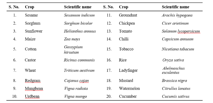
Twenty crop species were selected based on reported or suspected susceptibility to M. phaseolina. The list (common name and scientific name) is presented in Table 1.
Seeds were surface-sterilized with 1 per cent sodium hypochlorite (NaOCl) for 1-2 min, rinsed three times with sterile distilled water and blotted dry on sterile filter paper prior to inoculation.
PDA plates were inoculated centrally with a 5 mm disc of five-day-old M. phaseolina culture and incubated at 30 ± 1 °C for 48 h to allow pre-colonization. Ten surface-sterilized seeds of each species were placed equidistantly around the fungal colony on each plate. Four replicate plates per species (total n = 40 seeds per species) were maintained. To aid seed germination, 2 ml sterile distilled water was added to each plate prior to incubation. Plates were incubated in complete darkness at 30 ± 1 °C for six days (Manici et al., 1995; Beas-Fernandez et al., 2006).
On day seven, seed and seedling responses were assessed using a 0–5 scale (Manici et al., 1995; Cervantes-Garcia et al., 2003) where: 0 = healthy seed and seedling; 1 = discoloration of seed portion in contact with mycelium; 2 = seed integuments colonized by mycelium and microsclerotia but seedling healthy; 3 = seed coat unaffected but seedling infected; 4 = both seed integuments and seedling infected; 5 = seed infected and failed to germinate.
Virulence Index (VI) for each species was calculated following Beas-Fernandez et al. (2006):

Mean VI values were computed for each species. Standard error of mean (SEm ±), critical difference (CD at P = 0.05) and coefficient of variation (CV) were calculated.
In vitro screening revealed marked variation in M. phaseolina affinity among the 20 crops tested. Based on calculated Virulence Indices, crops were classified into three groups: Highly virulent (VI 4.6-4.9), Moderately virulent (VI 3.0-4.4) and Low virulent (VI 2.2-2.6). The summary of results is presented in Table 2.
Several pulses and oilseeds like sesame, groundnut, green gram, chickpea, redgram and black gram along with maize and brassica and cucurbits displayed the highest virulence (VI 4.6-4.9). Most of these crops exhibited seed integument invasion and seedling infection or failure to germinate (scores 4-5). This strong affinity corroborates with earlier reports that M. phaseolina is particularly aggressive on pulses and oilseeds under favorable conditions (e.g., warm, dry environment) and is capable of causing severe seedling blight and establishment failure. Production of cell wall-degrading enzymes (CWDEs) and phytotoxins likely contribute to rapid tissue maceration and seed death in these hosts (Ramos et al., 2016; Degani et al., 2023).
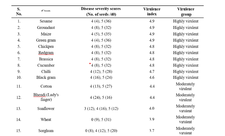
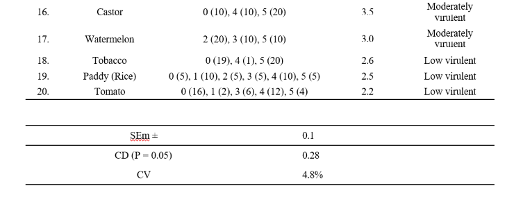
Cotton, bhendi, sunflower, wheat, sorghum, castor and watermelon fell into the moderate virulence category (VI 3.0-4.4). Symptoms were often less severe with partial seed coat invasion or seedling infection suggesting partial tolerance or structural/biochemical defenses reducing pathogen progress. Differences in host cell wall composition, seed coat thickness or preformed inhibitors may account for reduced colonization in these crops (Kaur et al., 2019; Hameed et al., 2022).
Tobacco, rice and tomato exhibited the lowest virulence indices (VI 2.2-2.6), generally showing discoloration at the seed portion in contact with mycelium or limited seedling symptoms (scores 1-2). Such low susceptibility could reflect effective early defense responses (hormonal signaling involving salicylic acid, jasmonic acid or ethylene) or physical barriers that limit fungal ingress (Radadiya et al., 2021). These crops may be useful models for dissecting resistance mechanisms and identifying candidate genes for breeding.
The pronounced host variation noted here is consistent with the recognized isolate-dependent pathogenicity of M. phaseolina. Reports from diverse regions indicate variability in aggressiveness on specific hosts (Rathore et al., 2024; Lokesh et al., 2020). Similarity between studies for example, reports of limited maize infection by some isolates versus high virulence on maize in the present study underline the influence of isolate genotype, host genotype and experimental conditions on observed pathogenicity (Ramos et al., 2016; Rathore et al., 2024).
High VI values for economically important crops such as sesame and groundnut underscore the need for integrated management. Due to longevity of microsclerotia in soil, crop rotation alone may reduce but not eliminate inoculum. Biological control agents (e.g., Trichoderma spp., Bacillus spp.) and the deployment of resistant genotypes appear promising components of integrated strategies (Khamari and Hashmi, 2019; Singh et al., 2022). Crops categorized in the low virulence group may possess effective defense mechanisms and further investigation of these mechanisms could provide useful leads for breeding durable resistance.
In vitro affinity assay demonstrated that Macrophomina phaseolina shows high affinity for pulses and oilseeds (VI 4.6-4.9), moderate affinity for cereals and cucurbits (VI 3.0-4.4) and low affinity on tobacco, rice and tomato (VI 2.2-2.6) under the tested conditions. These results highlight the broad host range and aggressive potential of M. phaseolina, particularly on pulses and oilseeds, and provide a basis for prioritizing crops for management, surveillance and breeding for resistance.
Beas-Fernandez, R., Manici, L.M and Cervantes-Garcia, 2006. Virulence variability of Macrophomina phaseolina isolates from different hosts and soil types. Phytopathologia Mediterranea. 45(2): 127- 136.
Cervantes-Garcia, D., Manici, L.M and Singh, S.D. 2003. Interaction of Macrophomina phaseolina with seeds of different host plants under controlled conditions. Journal of Mycopathological Research. 41(1): 37-42.
Degani, O., Dor, S and Drori, R. 2023. Global diversity and pathogenic potential of Macrophomina phaseolina isolates from major crops. Frontiers in Plant Science. 14: 115672.
Hameed, A., Niazi, M., Iqbal, S and Haider, S. 2022. Physiological and biochemical responses of oilseed crops to Macrophomina phaseolina infection. Journal of Oilseed Research. 39(1): 56-63.
Kaur, R., Singh, J., Kaur, P and Brar, H.S. 2019. Structural and biochemical defense mechanisms in cereals against Macrophomina phaseolina. Indian Phytopathology. 72(1): 87-94.
Khamari, R and Hashmi, A. 2019. Biological management of Macrophomina phaseolina causing dry root rot in groundnut. Indian Journal of Agricultural Research. 53(4): 456-462.
Lokesh, K., Maheshwari, R., Ramesh, K and Narayan, R. 2020. Assessment of yield losses due to dry root rot of chickpea caused by Macrophomina phaseolina. Legume Research. 43(2): 206-212.
Manici, L.M., Bonora, P and Caputo, F. 1995. Pathogenicity of Macrophomina phaseolina and its interaction with other soilborne fungi in chickpea. Plant Pathology. 44(4): 675-683.
Radadiya, C., Patel, D and Sharma, K. 2021. Hormonal defense pathways and induced resistance against necrotrophic pathogens in tomato. Journal of Plant Protection Sciences. 13(2): 89-98.
Ramos, B., Marquez, N and Giordano, A. 2016. Production of cell wall degrading enzymes by Macrophomina phaseolina isolates from different hosts. Mycopathologia. 181(5–6): 387-396.
Rathore, S., Saini, R and Mehta, R. 2024. Isolate-dependent variability of Macrophomina phaseolina infecting maize under subtropical conditions. Journal of Plant Disease Research. 39(1): 12-18.
Singh, P., Sharma, N and Reddy, K.R. 2022. Evaluation of biocontrol agents and resistant genotypes against Macrophomina phaseolina in sesame under field conditions. Indian Phytopathology. 75(2): 345-352.