

0 Views
Department of Plant Pathology, ANGRAU-S.V. Agricultural College, Tirupati-517 502.
Xanthomonas oryzae pv. oryzae (Xoo), the causal agent of bacterial leaf blight (BLB) in rice, poses a significant threat to rice cultivation in many parts of the world. The present study aimed to isolate and identify Xoo from infected rice leaves and confirm its pathogenicity, followed by preliminary biochemical and molecular characterization. Pathogenicity of the isolates was confirmed by clip inoculation on BPT 5204 cultivar, which reproduced BLB symptoms within 14 days. Biochemical characterization of the three isolates revealed catalase positivity, indicating the production of the catalase enzyme capable of breaking down hydrogen peroxide. The KOH solubility test yielded a positive result, supporting the Gram-negative nature of the isolates. However, the oxidase test was negative, which aligns with typical features of Xoo. Molecular characterization of three isolates was done with Xoo specific primers which yielded 290 bp size amplicon. These findings confirm the easy and reliable way to confirm the pathogen as Xoo without further sequencing.
KEYWORDS: Catalase, molecular characterization, oxidase, pathogenicity, Xanthomonas.
Xanthomonas oryzae pv. oryzae (Xoo) is a major bacterial pathogen causing bacterial leaf blight in rice, significantly impacting global rice production (Mondal et al., 2019; Zhao et al., 2007). The pathogen can cause yield losses of 10-50 per cent and is prevalent in most rice-growing countries (Ayusma et al., 2024; Yashitola et al., 2000). The bacterium can survive for extended periods and transmit from seed to plant to seed, enabling its persistence across growing seasons (Mondal et al., 2019).
Recent studies have focused on developing effective control measures against Xoo. These include the use of essential oils like eucalyptus and citronella (Ayusma et al., 2024), antimicrobial peptides such as melittin (Shi et al., 2016), and the development of highly sensitive real-time detection methods (Zhao et al., 2007). Genomic studies of Xoo strains from diverse geographical locations and altitudes are also contributing to a better understanding of its virulence factors and host-pathogen interactions (Asysyuura et al., 2017), paving the way for more targeted and effective management strategies for this significant rice pathogen.
Bacterial leaf blight (BLB) of rice, caused by Xanthomonas oryzae pv. oryzae (Xoo), manifests as severe symptoms that can lead to significant crop losses.
X. oryzae pv. oryzae enters the plant through wounds or hydathodes on the leaf margin and subsequently produces systemic infection (Mew, 1987). The disease could be characterized mainly into two distinct phases: the “leaf blight phase” and the “kresek phase.” The kresek phase occurs immediately after transplantation and is destructive (Reddy and Ou, 1976; Ou, 1985). The primary symptom is leaf wilting (Kim et al., 2015). The wilting is often accompanied by yellowing and drying of the leaves, resulting in characteristic blight symptoms. (Babu et al., 2003). The pathogen primarily colonizes and multiplies in the xylem vessels of rice plants, causing significant damage to the host tissue (Noda & Kaku, 1999).
The severity of symptoms can vary depending on the strain of Xoo and the rice cultivar’s resistance. The incidence and progression of diseases are heavily influenced by topography and climate. Specific amounts of sunlight and dryness impact the development of the causal agent. The disease commonly occurs in regions where monthly rainfall exceeds 200mm. Lesion formation tends to occur at temperatures ranging from 25 to 30°C. In the early stages, acidic soil is believed to significantly contribute to the disease (Maruyama, 1909). Heavy use of fertilizers is a key agricultural practice that impacts the onset of bacterial blight in rice. Among the three primary fertilizer components,
nitrogen is primarily responsible for promoting disease occurrence (Cha, 1982). The density of rice planting also plays a role in the disease’s severity, with greater severity observed under wider spacing while maintaining a constant nitrogen level. Colonies of Xoo appear as shiny, pin headed, mucoid, yellow and convex on modified Wakimoto media (Shankara et al., 2016).
Various methods have been used in the identification of the pathogen such as biochemical tests (Cruz et al., 1984), serological assays (Benedict et al., 1989), fatty acids and metabolic profiling (Chase et al., 1992; Merieux and Balme-les Grottes, 1993)
The preliminary diagnosis of Xoo lay the groundwork for future studies on virulence, race differentiation, and biocontrol strategies to manage BLB in rice.
Leaf samples showing water-soaked to yellowish orange lesions along the leaf margin were collected from three districts of Andhra Pradesh viz., Kurnool, Tirupati and Nellore were brought to lab for isolation of the pathogen and the isolates were named as XooK, XooT, XooN.
Infected leaves were excised with sterile scalpel. The leaf bits were sterilized with 1 per cent sodium hydroxide for three minutes and then washed with sterile distilled water. The leaf bits were macerated in sterile water to allow the bacteria to ooze out from the leaf tissue. After the water turns turbid, a loopful of suspension was streaked on modified Wakimoto Agar medium (MWA). After 48 hrs of incubation the emerging colonies which appeared as shiny, mucoid, convex, yellow colour were subcultured onto MWA media. The cultures were maintained on slants at 4oC in refrigerator for short term use and in 20 per cent glycerol at -20°C for long term preservation.
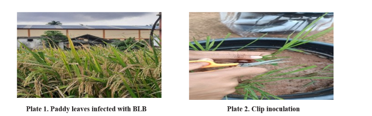
Artificial inoculation of the three Xoo isolates viz XooK, XooT, XooN was carried out using the leaf clip inoculation technique to prove pathogenicity under greenhouse conditions. Studies were conducted on BPT 5204 cultivar with 48h old culture. The bacterial suspension was clip inoculated to 45 days old plants by excising 2-3 cm of rice leaves with sterilized scissors immersed in the bacterial suspension (108 cells/ml). After inoculation plants were covered with polythene bags to maintain humid conditions. Plants were observed for symptoms after 72 h. The pathogen was re-isolated from artificially inoculated plants to prove Koch’s postulates and obtained bacterial colonies similar to the original culture.
Biochemical Characterization of Xanthomonas oryzae pv oryzae
Bacteria were heat fixed on a glass slide and was treated with (0.5%) crystal violet for 30 seconds then washed with running tap water. Then decolorized with (95%) ethanol for 30 seconds, washed again and counter-stained with safranin for 1 min. It was eventually washed with water and observed under microscope (Jonit et al., 2016).
KOH test was performed to confirm the gram staining results. The bacterial were aseptically transferred with a sterile loop onto the glass slide containing a drop of 3 per cent KOH solution and stirred for 10 seconds using a quick circular motion of hand (Suslow et al., 1982).
A loopful of 24 h old culture was smeared on a glass slide and few drops of hydrogen peroxide was added in the smear. The reaction is positive if gas bubbles are produced (Taylor and Achanzar, 1972).
Xoo was cultured on nutrient agar, supplemented with 1 per cent glucose. A loopful of 24 h was rubbed on filter paper impregnated with 15 per cent (w/v) freshly prepared aqueous solution of kovacs oxidase reagent (Kovacs, 1956). A colour change to purple is considered as positive.
The genomic DNA of three isolates was extracted by suspending a loopful of bacteria in TE buffer (500µl) followed by addition of 6 µl of Proteinase
K and 5 µl of RNAse A and incubated at 65°C for 30 minutes. NaCl (5M, 100 µl) and CTAB (80 µl) were added and once again incubated at 65°C for 30 minutes. An equal volume of chloroform: isoamyl alcohol (24:1) was added, mixed well and centrifuged at 13,000 rpm for 15 min. The supernatant was transferred to a new microcentrifuge tube. Chilled isopropanol was added and incubated overnight at -20°C. The next day the tube was centrifuged at 10000 rpm for 10 min, and supernatant was discarded to collect DNA pellet. Ethanol (500µl) was added, centrifuged and supernatant was discarded and pellet was air dried. Finally the DNA was dissolved in 30 µl of molecular-grade water and stored at -20°C.
DNA was quantified using Nanodrop. The molecular characterization was done using X00290 F/R specific primers of Xoo. For amplification of DNA using specific primer the PCR conditions include initial denaturation at 94°C for 5 minutes, 25 cycles of denaturation at 94°C for 60 seconds, annealing at 56°C for 30 seconds followed by extension at 72°C for 60 seconds, final extension at 72°C 10 minutes and reaction was kept hold at 4°C. The PCR products were analyzed on 1 per cent agarose gel electrophoresis and amplified DNA was viewed under the UV transilluminator.
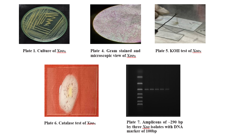
After 48 hours of incubation, small, pin-headed, raised, shiny, mucoid, convex yellow colonies were identified as Xoo. Single colonies were picked and transferred to modified Wakimoto Agar media for obtaining pure cultures of Xoo (Plate 3) Similarly, Shankara et al. (2016) isolated Xoo and reported that
colonies are mucoid and slightly convexed.
Koch’s postulates were followed to prove the pathogenic nature of Xanthomonas oryzae pv. oryzae. The three Xoo isolates were subjected to pathogenicity testing by leaf clipping method. Symptoms of BLB were observed on rice leaves after seven days of inoculation. Initially small water soaked lesions were observed which after fourteen days developed as stripes from clipped end forming yellow-brown lesions with wavy margins. Reisolation was done from these lesions and compared to original culture to confirm pathogen’s identity.
Similarly Akhtar et al. (2008) and Chandraprakash et al. (2018) performed isolation and pathogenicity tests and reported the water soaked lesions appeared in 5-7 days after inoculation of pathogen.
The three isolates exhibited Gram negative reaction with red colour when studied under light microscope. Since Gram negative bacteria have thin peptidoglycan layer, they do not retain purple colour and are counter stained by safranin giving red colour appearance as seen in Plate3.
Potassium hydroxide (KOH) test is an excellent validation assay for Gram staining (Suslow et al., 1982). The cell walls of Gram-negative bacteria are broken down in the presence of KOH and this results in releasing of the viscid chromosomal material which causes the bacterial suspension to become thick and stringy. The three Xoo isolates produced a characteristic slimy thread like structure when treated with 3 per cent KOH confirming gram-negative nature of Xoo(Plate 4). Rukhsana et al. (2012), reported that Xoo was Gram
negative, rod shaped and produced red colour when counter stained with safranin.
All the isolates were found to be positive for catalase production. Bubbles were formed when few drops of hydrogen peroxide were added on bacteria smear (Plate 6.) It is because catalase enzyme is able to decompose hydrogen peroxide releasing oxygen and water. Catalase plays a crucial role in the defense mechanisms of Xoo against oxidative stress, particularly in detoxifying hydrogen peroxide (H2O2) (Pan et al., 2016).
The isolates when smeared on filter paper dipped in Kovacs reagent there was no colour change indicating the isolates are negative for oxidase test. Similar results were obtained by Islam et al., 2023 for oxidase test. It may be because the bacteria may not be able to produce cytochrome c oxidase, an enzyme in the electron transport chain.
The 16S rDNA primers 27F/1492R of three isolates of Xoo yielded amplicons of ~1500bp and when the PCR was done with specific primer set XOO290F/R, ~290 bp size amplicons were obtained. These results are in corroboration with studies of Tuan et al., 2017. With the obtained amplicons with specific primers the three bacterial isolates were considered as Xanthomonas oryzae pv oryzae.
The present study successfully isolated and identified Xanthomonas oryzae pv. oryzae (Xoo), the causal agent of bacterial leaf blight (BLB) in rice. The isolates displayed characteristic yellow, mucoid colonies on Wakimoto Agar and reproduced typical BLB symptoms through clip inoculation. Biochemical tests confirmed the Gram-negative nature, catalase activity, and oxidase negativity of the isolates traits consistent with Xoo. Molecular confirmation using Xoo-specific primers resulted in distinct amplicons of ~290 bp validating the identity of the isolates. These findings offer a reliable and easy protocol for preliminary diagnosis of Xoo by usng specic primers without going for sequencing and lay the groundwork for future studies on virulence, race differentiation, and biocontrol strategies to manage BLB in rice.
Akhtar, M.A., Rafi, A and Hameed, A. 2008. Comparison of methods of inoculation of Xanthomonas oryzae pv. oryzae in rice cultivars. Pakistan Journal of Botany. 40(5): 2171-2175.
Aldrick, S.J., Buddenhagen, I.W., Reddy, A.P.K. 1973. The occurrence of bacterial leaf blight in wild and cultivated rice in northern Australia. Australian Journal of Agricultural Research. 24: 219–227.
Ayusma, A.N., Widiastuti, A and Arwiyanto, T. 2024. Potency of Eucalyptus Oil and Citronella Oil in Suppressing Virulence Factors of Xanthomonas oryzae pv. oryzae. Jurnal Fitopatologi Indonesia. 20(4): 195-203.
Babu, R.M., Sajeena, A., Samundeeswari, A.V., Sreedhar, A., Vidhyasekeran, P and Reddy, M.S. 2003. Induction of bacterial blight (X. oryzae pv. oryzae) resistance in rice by treatment with acibenzolar S methyl. Annual Applied Biology. 143: 333-340
Benedict, A.A., Alvarez, A.M., Berestecky, J., Imanaka, W., Mizumoto, C.Y., Pollard, L.W., Mew, T.W., Gonzalez, C.F. 1989. Pathovar-specific monoclonal antibodies for Xanthomonas campestris pv. oryzae and for Xanthomonas campestris pv. oryzicola. Phytopathology. 79: 322–328.
Cha, K.H., Kim Y.S., Kim, H.J., Lee, D.K and Kim, M.S. 1982. Effects of application levels of fertilizer on the susceptibility to bacterial leaf blight, yield and quality of grains in nineteen rice cultivars in Jeonnam Region. Korean Journal of Plant Protection. 21(4): 216-221.
Chandraprakash, S., Manonmani, K., Revathy, N and Devi, E.K., 2018. Management of bacterial leaf blight disease of paddy (Oryza sativa) caused by Xanthomonas oryzae pv. oryzae using bacteriophages. International Journal of Current Microbiology and Applied Sciences. 7: 3066-3080.
Chase, A.R., Stall, R.E., Hodge, N.C., Jones, J.B. 1992. Characterization of Xanthomonas campestris strains from aroids using physiological, pathological, and fatty acid analyses. Phytopathology 82: 754-759.
Cowan, S.T. 1974. Manual for the identification of medical bacteria. Cambridge University Great Britain Journals Press. 238.
Cruz, C.V., Gossele, F., Kersters, K., Segers, P., Van den Mooter, M., Swings, J., De Ley, J. 1984. Differentiation between Xanthomonas campestris pv. oryzae, Xanthomonas campestris pv. oryzicola and the bacterial ‘brown blotch’ pathogen on rice by numerical analysis of phenotypic features and protein gel electrophoregrams. Microbiology 130: 2983– 2999.
Islam, M.A., Rahman, M.A., Akter, T., Islam, M.M., Salam, M.A., Misty, S.S., Hossain, M.A. and Haque, M.A. 2023. Collection, Pathotype Profiling, and Virulence Analysis of Xanthomonas oryzae pv. oryzae Causing Bacterial Leaf Blight Disease of Rice in Bangladesh. International Journal of Innovative Science and Research Technology. 8(3): 2456-2165.
Jonit, N.Q., Low, Y.C and Tan, G.H. 2016. Xanthomonas oryzae pv. oryzae, biochemical tests, rice (Oryza sativa), Bacterial Leaf Blight (BLB) disease, Sekinchan. Applied and Environmental Microbiology. 4: 63-69.
Kim, S. M., Suh, J. P., Qin, Y., Noh, T. H., Reinke, R. F and Jena, K.K. 2015. Identification and fine mapping of a new resistance gene, Xa40, conferring resistance to bacterial blight races in rice (Oryza sativa L.). Theoretical and Applied Genetics. 7(2):12-15.
Kovacs, N. 1956. Identification of Pseudomonas pyocyanea by the oxidase reaction. Nature. 178: 703.
Maruyama, H. 1909. Soybean cake application and leaf blight disease of rice. Shizuoka-ken Nokaiho. 142: 28-29.
Merieux, B., Balme-les Grottes, L. 1993. Evaluation of the Biolog GN MicroPlate system for identification of some plant-pathogenic bacteria. Plant Disease: 553–558.
Mew, T.W. 1987. Current status and future prospects of research on bacterial blight of rice. Annual Review of Phytopathology. 25(1): 359 – 382.
Mondal, S., Hossien, M.E., Akter, M.A., Haque, M.M., Ali, M.A and Islam, M.R., 2019. Survival and transmission of Xanthomonas oryzae pv. oryzae in rice seeds. Fundamental and Applied Agriculture. 4(1): 680-688.
Noda, T., & Kaku, H. (1999). Growth of Xanthomonas oryzae pv. oryzae In Planta and in Guttation Fluid of Rice. Japanese Journal of Phytopathology, 65(1), 9–14.
Ou, S.H. 1985. Rice diseases. 2nd edition Common wealth Mycological Institute. Kew, UK. 109-201.
Pan, X., Wu, J., Duan, Y., Xu, S. and Zhou, M. 2016. CatB is critical for total catalase activity and reduces bactericidal effects of phenazine-1-carboxylic acid on Xanthomonas oryzae pv. oryzae and X. oryzae pv. oryzicola. Phytopathology. 107(2): 163–172.
Reddy, C.R. and Ou, S.H. 1976. Characterization of Xanthomonas oryzae (Uyeda et. Ishiyama) Dowson, the bacterial blight pathogen of rice. Annual Review of Phytopathology. 42: 124-130.
Rukhsana, J., Thehreema, I and Huma, B. 2012. Isolation, characterization, preservation and pathogenicity test of Xanthomonas oryzae pv. oryzae causing BLB disease in rice. Pakisthan Journal of Botany. 44(1): 261 – 265.
Sanya, D.R.A., Onesime, D., Rohr, J.R., Ahohuendo, B.C., Syed-Ab-Rahman, S.F., Jia, A and Kim, K.M. 2022. A review of approaches to control bacterial leaf blight in rice. World Journal of Microbiology and Biotechnology. 38(7).
Shankara, K., Patil, M.B., Pramesh, D and Sunkad, G. 2016. Isolation and Characterization of Bacterial Leaf Blight of Rice (Xanthomonas oryzae pv. oryzae) Isolates from Southern India. Advances in Life Sciences. 5(14): 5625-5633.
Suslow, T.W., Schrotl, N and Isaha, M. 1982. Application of a rapid method for Gram differentiation of plant pathogenic bacteria without staining. Phytopathology. 72: 917-918.
Taylor, W.I and Achanzar, D. 1972, Catalase test as an aid to the identification of Enterobacteriaceae. Journal of Applied Microbiology. 24: 58 – 61.
Tran, T.T., Pérez-Quintero, A.L., Wonni, I., Carpenter, S.C., Yu, Y., Wang, L., Leach, J.E., Verdier, V., Cunnac, S., Bogdanove, A.J and Koebnik, R. 2018. Functional analysis of African Xanthomonas oryzae pv. oryzae TALomes reveals a new susceptibility gene in bacterial leaf blight of rice. PLoS Pathogens. 14(6): 1007092.
Tuan, T.Q., Hao, L.T and Khoa, N.D. 2017. Population diversity of Xanthomonas oryzae pv. oryzae causing bacterial leaf blight in rice fields of Can Tho. Journal of Biotechnology. 15(4). 753-761.
Yashitola, J., Reddy, A.P.K and Sonti, R.V. 2000. A widely distributed lineage of Xanthomonas oryzae pv. oryzae in India may have come from native wild rice. Plant disease. 84(4): 465-469.
Zhao, W.J., Zhu, S.F., Liao, X.L., Chen, H.Y and Tan,
T.W. 2007. Detection of Xanthomonas oryzae pv. oryzae in seeds using a specific TaqMan probe. Molecular Biotechnology. 35: 119-127.