
0 Views
Department of Plant Pathology, S.V. Agricultural College, ANGRAU, Tirupati-517502.
Brinjal little leaf (BLL) is a prevalent phytoplasma disease in India that induces severe economic losses every year. During
2021, symptoms of little leaf and witches’ broom were observed on brinjal plants grown in the horticultural fields of S. V. Agri- cultural College, Tirupati. Four symptomatic and two symptomless plants collected from the surveyed places were subjected to genomic DNA extraction and used as template in nested PCR assays with universal phytoplasma 16S rRNA primers, P1/P7 and R16F2n/R16R2. Amplification of ~1.25 kb product was obtained only from symptomatic BLL plants and the positive control, but not from any asymptomatic plants.
KEYWORDS: Eggplant/Brinjal, 16S rRNA, Nested PCR assay.
Brinjal (Solanum melongena L.) is the most important vegetable cultivated all over the world. India is the second largest producer of brinjal in the world and Andhra Pradesh is a leading brinjal growing state of India (Anonymous, 2019). The crop is challenged by several diseases caused by fungi, bacteria, viruses and other insect pests of which brinjal little leaf (BLL) is one of the most important disease caused by phytoplasma causing considerable economic losses as the infected plant fails to produce a single fruit. Phytoplasma associated diseases in vegetable crops are prevalent in Asian, African, and American continents. So far twenty- eight vegetable species have been reported to be infected by different strains of phytoplasma all around the world (Kumari et al., 2019). Brinjal Little Leaf disease was first recorded by Thomas and Krishnaswami (1939) in India with 100% yield loss (Rao and Kumar, 2017). Five ribosomal groups (16SrI, 16SrII 16SrVI, 16SrIX and 16SrXII) were reported associated with BLL disease at global level (Kumari et al., 2019). The phytoplasma groups 16SrI and 16SrVI (subgroup 16SrVI-D) are the most dominant reported to be associated with BLL disease in eggplants showing little leaf, shortening of internodes, witches’ broom, phyllody, stunting and yellowing with necrosis symptoms in India (Kumar et al., 2017; Rao and Kumar, 2017). The phytoplasma disease of eggplant was reported to be widespread in the areas, where overlapping crop cycles and weeds ensure high populations of leafhoppers and provide natural reservoirs for the different strains of phytoplasma (Rao and Kumar, 2017; Rao, 2021). But limited reports are available from Andhra state of India. Since, most of the farmers in Andhra Pradesh are cultivating brinjal for commercial sale in local markets, an attempt was made to detect, phytoplasma presence associated with BLL in the Chittoor district of Andhra Pradesh.
During April 2021 survey was conducted in horticultural fields of S. V. Agricultural College, Tirupati, Chittoor district, Andhra Pradesh. The disease incidence of BLL was assessed by counting number of infected plants visually over healthy plants by randomly selecting 5 × 5 meter plot at each location. Moreover symptomatic leaf samples were collected in each surveyed location were brought to the laboratory along with two healthy leaf samples for further processing. DNA was extracted from the symptomatic and non-symptomatic brinjal samples using CTAB method.
The total genomic DNA was isolated from the leaves of healthy and symptomatic plants by following CTAB (Cetyl Trimethyl Ammonium Bromide) method of Murray and Thomson (1980) with some modifications. The modifications were made to improve the quality of DNA.
DNA was assessed for its purity and intactness using agarose gel electrophoresis.
Preparation of 1% Agarose Gel: 1 g of agarose was placed in a conical flask containing 100 ml 1X TAE buffer. The conical flask along with its contents was placed in an oven until agarose gets melted completely
and clear solution was formed and then the flask was taken out from the oven and allowed to cool. 3 µl of Ethidium Bromide (10 mg ml-1) was added to this 100 ml of agarose gel and mixed thoroughly. Later the solution was poured slowly into the gel casting tray which is pre- set with 0.5 mm combs, to avoid the formation of bubbles. After solidification the gel with casting tray was placed in gel tank and the comb was removed gently without disturbing the wells that formed upon solidification.
The NanoDrop spectrophotometer (model ND1000) was used to assess the quantity and quality of DNA employing the following procedure. Before initializing the NanoDrop Reader, the pedestal was cleaned with tissue paper to remove the dust particles. Then for initializing the instrument, 1-2 μl of distilled water was placed on the lower pedestal, closed the upper one and clicked on measure option. Then the pedestal was cleaned with tissue paper and 1.5 μl of 1X TE buffer was placed on lower pedestal and repeated the procedure for blank measurement. After that, 2 μl of DNA sample was placed to measure the quality and quantity at A260 nm and A280 nm to assess the purity of DNA. The process is repeated for all the DNA samples. A ratio of ~1.8 is generally accepted as “pure” for DNA; a ratio of ~2.0 is generally accepted as “pure” for RNA. If the ratio is lower in either case, it may indicate the presence of protein, phenol or other contaminants that absorb strongly at/or near 280 nm.
Normalization of DNA samples was done to equalize the concentration of all the samples to be used for PCR reaction. The purpose of normalization was to avoid erroneous analyses due to difference in the brightness of the bands obtained after electrophoresing the amplified PCR products. Normalization was done by diluting the DNA samples with sterile distilled water to their required dilution factor which in turn depends upon the initial concentration of DNA sample (from quantification readings) and the type of analysis done (markers used). After normalization of samples the concentration of DNA was 100 ng µl-1.
It was done by using the formula: N1V1 = N2V2 PCR Amplification of Phytoplasmas DNA Primers used for 16S rRNA gene
The extracted DNA from the plants and insects was amplified for 16S ribosomal DNA with phytoplasma specific universal primer pair P1/P7 (Deng and Hiruki, 1991; Schneider et al., 1995) followed by nested primer
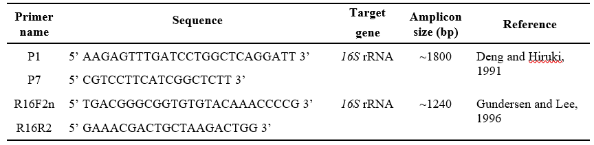
pair R16F2n/R16R2 (Gundersen and Lee, 1996) (Table 1).
PCR reactions were carried out in a Mastercycler (Eppendorf, Germany) and the cycling protocols for the amplification of different genes used are described herein.
First round of PCR assay for the conserved region of the 16S rRNA gene using P1/P7 primer pair and the cycling protocol used in the study are mentioned here under (Table 2).
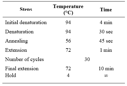
For nested PCR amplification of 16S rRNA using R16F2n/R16R2 primer pair and the cycling protocol used are mentioned in Table 3.
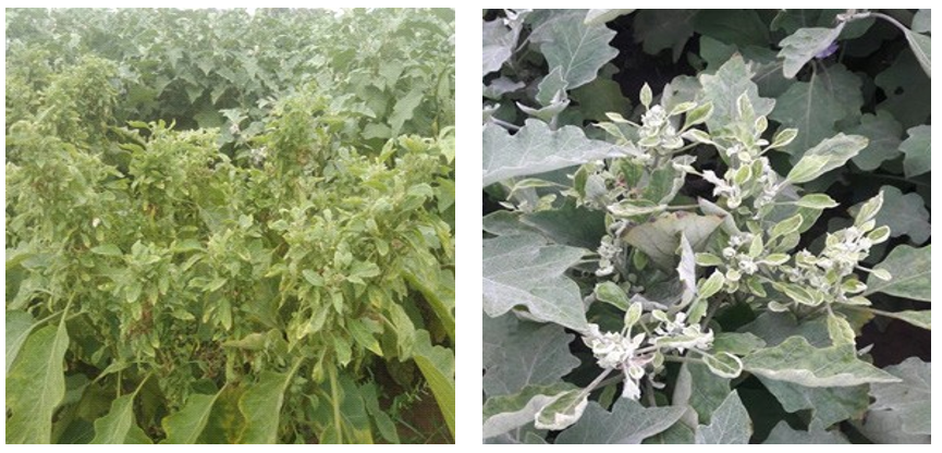
From all the collected samples, the affected brinjal plants showed the symptoms of littleleaf and witches’ broom (Figures 1a and 1b). The disease incidence was recorded from10% to 20%. The association of phytoplasmas in all the symptomatic samples was

confirmed by PCR amplification of ~1.8 kb products in the first round (Figure 2a). PCR and ~1.2 kb products in nested PCR assays with primer pair P1/P7 and R16F2n/R16R2, respectively, while no amplification was observed in the asymptomatic samples (Figure 2b) which confirmed the association of phytoplasma with symptomatic brinjal plants.
Phytoplasma belonging to 16SrII-D subgroup was found as the most widely distributed phytoplasma strain on other plant crops in India (Rao, 2021; Reddy et al., 2021) . Earlier the 16 srII- D phytoplasm were identified on brinjal from Uttar Pradesh and Karnataka (Kumar et al., 2017; Yadav et al., 2016) and 16SrVI-D (Azadvar and Baranwal, 2012; Kumar et al., 2017) was reported from various parts of India whereas, the presence of 16SrVI-D and 16SrI group in Bangladesh (Siddique et al., 2001), 16SrII-A in China (Cai et al., 2016), 16SrII-D in Oman (Al-Subhi et al., 2018) was reported in BLL diseased plants.
Naik et al. (2018) reported the association of 16SrVI-D sub-group of phytoplasma in Andhra Pradesh.

Lane M: 1kb ladder, Lane N: Negative control, Lane P: Positive control,
Lane 1: brinjal isolate sample-1 and Lane 2- Brinjal isolate sample-2
In the present study, the association of 16SrII-D from Andhra Pradesh was confirmed based on sequence comparison, phylogeny and RFLP analysis of 16S rDNA sequences. To our knowledge, this is the first report of the showing the association of 16SrII-D sub-group of phytoplasma with little leaf of brinjal in Andhra Pradesh. Recently, 16SrII-D subgroup phytoplasma was identified and characterized from chickpea and weeds from Kadapa, Andhra Pradesh (Reddy et al., 2021). In India, brinjal is cultivated in the same season with many other agricultural crops. The scenario of natural phytoplasma spread from brinjal to other plant species and vice versa, through an efficient vector species, is quite possible as reported in other states of India (Kumar et al., 2017).

This indicates that Brinjal Little Leaf disease is widely spreading and association of new sub-groups were reported based on the prevalence of alternate hosts and vectors in a particular locality. Keeping this in view, further studies on epidemiology along with possible natural spread sources and management are therefore a foremost need.
Al-Subhi, A.M., Hogenhout, S.A., Al-Yahyai, R.A and Al-Sadi, A.M. 2018. Detection, identification, and molecular characterization of the 16SrII-D phytoplasmas infecting vegetable and field crops in Oman. Plant Disease. 102: 576-588.
Anonymous 2019. http://www.mospi.gov.in/ [accessed 02.06.2021]
Azadvar, M and Baranwal, V.K. 2012. Multilocus sequence analysis of phytoplasma associated with brinjal little leaf disease and its detection in Hishimonas phycitis in India. Phytopathogenic Mollicutes. 2: 15-21.
Cai. H., Wang, L., Mu, W., Wan, Q., Wei, W., Davis, R.E., Chen, H and Zhao, Y. 2016. Multilocus genotyping of a ‘Candidatus Phytoplasma aurantifolia’-related strain associated with cauliflower phyllody disease in China. Annals of Applied Biology. 169. 64-74.
Deng, S and Hiruki, C. 1991. Amplification of 16S rRNA genes from culturable and nonculturable mollicutes. Journal of Microbiological Methods. 14: 53-61.
Gundersen, D.E and Lee, I.M. 1996. Ultrasensitive detection of phytoplasmas by nested-PCR assays using two universal primer pairs. Phytopathologia Mediterrenea. 35: 144-151.
Kumar, M., Madhupriya and Rao, G.P. 2017. Molecular characterization, vector identification and sources of phytoplasmas associated with brinjal little leaf disease in India. 3 Biotech. 7: 1-11.
Kumari, S., Nagendran, K., Rai, A.B., Singh, B., Rao,
G.P and Bertaccini, A. 2019. Global status of phytoplasma diseases in vegetable crops. Frontiers in microbiology. 10: 1349.
Murray, M.G and Thompson, W.F. 1980. Rapid isolation of high molecular weight plant DNA. Nucleic Acids Research. 8: 4321-5.
Naik, D.V.K., Reddy, B.V.B., Rani, J.S., Devi, S.J.R
and Prasad, K.V.H. 2018. Candidatus Phytoplasma trifolii associated with little leaf disease of Solanum melongena (Brinjal) in Andhra Pradesh, India. Journal of Pharmacognosy and Phytochemistry. 7: 3695-3697.
Rao, G.P. 2021. Our understanding about phytoplasma research scenario in India. Indian Phytopathology. 1-31.
Rao, G.P and Kumar, M. 2017. World status of phytoplasma diseases associate with eggplant. Crop Protection. 96: 22-29.
Reddy, M.G., Baranwal VK, Sagar, D and Rao, G.P. 2021. Molecular characterization of chickpea chlorotic dwarf virus and peanut witches’ broom phytoplasma associated with chickpea stunt disease and identification of new host crops and leafhopper vectors in India. 3 Biotech. 11(3): 1-23.
Schneider, B., Seemüller, E., Smart, C.D and Kirkpatrick, B.C. 1995. Phylogenetic classification of plant pathogenic mycoplasmalike organisms or phytoplasmas. In: Molecular and Diagnostic Procedures in Mycoplasmology. Vol. 2, pp 369–380. Eds S Razin and JG Tully. Academic Press,
Siddique, A.B.M., Agrawal, G.K., Alam, N and Reddy,M.K 2001. Electron microscopy and molecular characterization of phytoplasmas associated with little leaf disease of brinjal (Solanum melongena L.) and periwinkle (Catharanthus roseus) in Bangladesh. Journal of Phytopathology. 149: 237- 244.
Thomas, K.M and Krishnaswami, C.S. 1939. Little leaf—a transmissible disease of brinjal. Proceedings of the National Academy of Sciences. 10: 201-212.
Yadav, V., Mahadevakumar. S., Tejaswini, G.S., Shilpa, N., Amruthavalli and Janardhana, G.R. 2016. First report of 16SrII-D phytoplasma associated with eggplant big bud (Solanum melongena L.) in India. Plant Disease. 100: 517.